Prostate Zones and Prostate Cancer – All You Need to Know


Article Summary
- The prostate zones have clear interrelations with prostate carcinoma.
- Learning about the prostate gland and the human body’s anatomy can help men better understand their susceptibility to cancer.
- Based on the reports, most cancers begin in the PZ zone, whereas only a fraction of the tumors are found in fibromuscular Stroma.
Prostate carcinoma is a serious health problem for men all around the globe. In fact, the disease is affecting around 1 in every 6 men.
Those who suspect they have cancer do a prostate biopsy and MRI screening. The sooner you detect the illness, the easier it can be to manage it.
But, it’s critical to understand the prostate zones when you want to know more about them. Identifying the zone can help you learn more about the male body and its cancer susceptibility. This is a detailed review of the zones and their connection to prostate cancer.
What Is the Function of the Prostate?
The prostate has a key role to play in the male reproductive system. The prostate is a gland situated just under the bladder and in front of the rectum. It wraps around the prostatic urethra, which is the upper region of the urethra.
Its primary purpose is to create a prostatic fluid that is necessary for semen production. This fluid transports the sperm during orgasm. There are countless smooth muscle cells within the prostate gland. During ejaculation, the cells contract and press the fluid into the urethra.
The prostate gland is made of anterior, apex, base, posterior, and lateral surfaces. It is also divided into multiple lobes, such as the median lobe, anterior lobe, posterior lobe, and lateral lobe. Because of how close the prostate is to other sexual and urinary organs when it is not functioning properly, the prostate can interfere with sexual and urination function.
For example, when a man turns 40, their prostate gland tends to become bigger. The prostate volume also changes. Eventually, it will constrict the urethra, which, in turn, prevents or slows urine flow. This is what experts call benign prostatic hyperplasia (BPH), or previously known by the term benign prostatic hypertrophy.
The prostatic venous plexus is in charge of draining the internal iliac vein that is linked to the venous plexus. This is believed to be the route of the spread of cancer to the bones.
For the prostate gland to work properly, it needs adequate amounts of specific hormones, particularly testosterone. Since the prostate gland is made of various types of tissue, it is also divided into different zones. Understanding these zones can give you a clear insight into prostate cancer development.
Prostate Zones
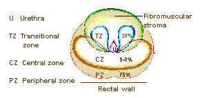
Researchers have divided the prostate into distinct zones based on their primary function. You have the Transitional zone, Central zone, and Peripheral zone. There is also the fibromuscular Stroma, which is considered the fourth zone for some experts. Prostate cancer can start to form and potentially even spread from any of these zones.
Image Source: https://training.seer.cancer.gov/images/prostate/zones.jpg
Unlike typical ultrasound, mpMRI (multiparametric magnetic resonance imaging) clearly depicts these zones. It can also help differentiate between diseased tissue and healthy prostate tissue.
Prostate MRI is used to detect prostate carcinoma in any of the zones, including extracapsular extension (tumor beyond or at the edge of prostate cancer). The ADC values (apparent diffusion coefficient) help measure water molecules’ magnitude within the affected tissue.
To find out more about these zones, check out the detailed overview below.
Transitional Zone (TZ)
The TZ takes up 5% of the adult gland and envelops the urethra that moves through the prostate, also known as the prostatic urethra.
From 20% to 30% of prostate cancers begin to form in this particular zone. This section may be small for a young adult, but the transitional zone also increases as the person grows older. We expect prostate enlargement.
In time, it will take up a larger percentage of the gland and can cause prostatic hyperplasia. Based on reports from 494 patients, a TZ tumor has better biochemical relapse-free survival than the one found in the peripheral zone. They also show a better outcome than their peripheral zone counterparts.
Central Zone (CZ)
The central zone features 20% of the adult gland. It is located around the ejaculatory duct, which runs to the prostatic urethra. Compared to other zones, only a fraction of prostate carcinomas begins here.
According to a clinical trial of 2,494 tumors, just 63 (2.5%) were found in the CZ. However, the tumor that grows in this region tends to become more aggressive.
Based on a different study of 211 patients, cancers associated with the central zone had a much higher Gleason score, seminal vesicle invasion, extracapsular extension, and prostate-specific antigen value compared to cancers in different zones. That’s why cancers involving CZ are related to an aggressive disease.
Peripheral Zone (PZ)
The Peripheral zone features most of the adult gland (70%). The back of the gland lies under the capsule and encloses the distal urethra. Around 70% to 80% of cancers begin to form in this zone. When a doctor does a DRE (digital rectal exam), they will feel the back of the gland.
Researchers believe the peripheral zone of the prostate is more susceptible to tumor development than the transitional zone. But, both zones have unique molecular differences that can play a fundamental role in the predisposition to the progression and development of prostate cancer.
DWI (diffusion-weighted imaging) measures the water molecules and Brownian motion in the voxel of tissue. According to a T2 weighted imaging report of the PZ, the prostate appears to be an area of high signal intensity. While the central gland features a variable signal intensity. Prostate carcinoma in the PZ can seem like an area of hypo-intense signal intensity.
Fibromuscular Stroma
Located anteriorly in the gland, the fibromuscular Stroma integrates with the tissue of the urogenital diaphragm. This particular area doesn’t have any gland. Instead, the prostatic tissue is made of glandular and connective tissue.
The anterior fibromuscular Stroma is responsible for forming the complete anterior surface of the prostate gland. It basically functions as a thick apron that shields the glandular tissue regions from view.
According to medical reports, the anterior fibromuscular Stroma is a group of fibromuscular tissue closer to the front of the TZ. The anterior fibromuscular Stroma is typically hypovascular, probably because of its fibrous nature. Rarely any cancers are in this zone.
Why Are These Zones Important?
Plenty of research shows that prostate carcinomas exhibit unique features based on the zone they form in. Reports from Stanford University indicate there are noteworthy biological differences between prostate carcinoma in the peripheral and transitional zone.
Scientists evaluated 1,354 prostate carcinoma patients who had a radical prostatectomy. They analyzed each cancer based on the zonal location. Experts found that:
- People with transitional zone cancer had a lower risk of cancer coming back.
- People with peripheral zone cancer had a high incidence of chronic and acute inflammation in PZ compartments.
- Patients with transitional zone cancer had a reduced possibility of extracapsular extension and seminal vesicle invasion.
- People with transitional zone cancer had increased total cancer volume and pre-operative PSA values compared to peripheral zone cancers. But a better recurrence-free survival rate.
Identifying Signs of Cancer in the Prostate
Prostate carcinoma is the second most widespread cancer in men and the 5th cause of death around the globe. In 2018, more than 1 million new cases of prostate carcinoma were recorded worldwide. In its early stages, cancer can be asymptomatic and only require active surveillance.
But, when it starts to cause problems and get in the way of your daily life, that’s when the cancer is easy to recognize. The body goes through a series of changes that could be a red flag for a more serious health complication. Patients need to do a serum PSA test to screen for cancer.
Here are some of the most typical signs to identify prostate carcinoma.
- Blood in the urine
- Need to urinate at night frequently
- Need to urinate over and over again
- Recent erectile dysfunction
- Burning or painful sensations when urinating
- Pain and discomfort when sitting
Other non-cancerous prostate illnesses, like enlarged prostate and stromal BPH, can trigger similar symptoms. Although BPH doesn’t boost cancer risk, the malignant BPH nodules can be a sign of prostate carcinoma.
However, the symptoms listed here could also be due to another medical condition completely unrelated to cancer. It could be a urinary symptom from a bladder condition.
In the case of a BPH, three-dimensional transrectal ultrasound can be used to identify the zones’ structural differences. It can be performed prior to a biopsy of the prostate gland.
If cancer spreads towards the surrounding tissue or organs, it could cause signs such as:
- Exhaustion
- Feet or leg swelling/fluid buildup
- Pain in the shoulders, thighs, hips, back, or other bones
- Bowel habit changes
MRI is the main imaging modality for evaluating the state of the prostate gland. The MRI can detect the stage of cancer and assess the disease. It could also analyze the possible invasion to other structures, such as the external urethral sphincter, rectum, pelvic wall, bladder, or levator ani muscles.
Therefore, understanding the MR imaging of the gland and pelvic structures can help interpret the cancerous or non-cancerous condition.
If you are worried about any of the signs you experience, consult with a doctor as soon as possible. They can help identify the problem and help soothe the symptoms. With proper testing, they can give you a diagnosis and suggest the best form of treatment. The treatment will vary according to the patient’s cancer stage.
In prostate carcinoma, the presence of metastasis to the pelvic lymph node is believed to represent a systematic illness. It also links with poor diagnoses post radical prostatectomy. As a result, other treatments, like hormone therapy instead of surgery, are often a recommendation.
What’s the Survival Rate for This Type of Cancer?
In most men, the 5-year survival rate from prostate carcinoma is almost 100%. But, if the condition has spread outside the prostate gland and affects neighboring parts of the human body, the rate can drop to 31%.
That’s why for some tumors, early screening is critical. Doctors can spot the illness in its early phases and manage it before it becomes a real threat. Plus, the sooner you detect the problem, the easier it can be to manage. You could only detect prostate carcinoma early with PSA testing.
So, those at risk or have someone in the family who has developed the condition may benefit from regular screening. But, before you do any tests, you must consult with a doctor. They will let you know the best approach for your condition and whether or not you need screening and testing.
Remember, everyone’s body is different. Even if the body is vulnerable to carcinoma, that doesn’t mean you should expose the system to constant screening. You should only test for carcinoma if your doctor believes it is the right approach.
Final Thoughts
The prostate zones have clear interrelations with prostate carcinoma. Learning about the prostate gland and the human body’s anatomy can help men better understand their susceptibility to cancer. Based on the reports, most cancers begin in the PZ zone, whereas only a fraction of the tumors are in fibromuscular Stroma.
The information listed here can help you get a detailed guideline of everything that could happen within these zones. If you think you are at risk of developing this type of cancer, it is best to consult a urologist. The doctor will assess the prostate gland via biopsy and imaging tests to see what’s happening in your body.
This article is for informational purposes only and does not serve as medical advice. The details provided here are not a replacement for, and should never be depended upon as, professional medical advice. Always consult your physician regarding the potential risks and benefits of any treatment.
Our Medical Review Process
At Ben’s Natural Health, we are committed to maintaining the highest standards of accuracy, transparency, and scientific integrity. Every piece of content is carefully developed by medical professionals and undergoes a thorough review every 12 to 24 months. This ensures that our information remains current, reliable, and rooted in credible, evidence-based research. We reference only peer-reviewed studies from reputable medical journals, providing full citations and direct links to enhance trust and confidence. Learn more about our medical review process and research standards.
Our Editorial Guidelines
For over 25 years, Ben’s Natural Health has been a trusted source of scientifically backed, reliable health information. Our editorial guidelines uphold the highest quality and integrity for every article we publish. Each piece is written by qualified experts and undergoes independent quality checks. We prioritize transparency by clearly displaying contributor credentials and biographies at the beginning of every article. Read more about our editorial standards.
Medical Disclaimer
The content on this blog is for informational purposes only and should not be considered a substitute for professional medical advice, diagnosis, or treatment. While our articles are authored and reviewed by licensed medical professionals, they may not address your specific health concerns. Always consult a qualified healthcare provider before making any medical decisions.
Article Sources
- National Cancer Institute. Zones of the Prostate. Retrieved from: https://training.seer.cancer.gov/prostate/anatomy/zones.html
- Jennifer A. Sinnott. (2015). Molecular differences in transition zone and peripheral zone prostate tumors. National Institutes of Health. Retrieved from: https://www.ncbi.nlm.nih.gov/pmc/articles/PMC4572920/
- Christopher R King. (2008). Prognostic significance of prostate cancer originating from the transition zone. National Library of Medicine. Retrieved from: https://pubmed.ncbi.nlm.nih.gov/18799332/
- Ronald J. Cohen. (2008). Central Zone Carcinoma of the Prostate Gland: A Distinct Tumor Type With Poor Prognostic Features. AHA Journals. Retrieved from: https://www.auajournals.org/doi/10.1016/j.juro.2008.01.017
- Hebert Alberto Vargas. (2012). Normal central zone of the prostate and central zone involvement by prostate cancer: clinical and MR imaging implications. National Library of Medicine. Retrieved from: https://pubmed.ncbi.nlm.nih.gov/22357889/
- Zaki Shaikhibrahim. (2011). The peripheral zone of the prostate is more prone to tumor development than the transitional zone: is the ETS family the key?. National Library of Medicine. Retrieved from: https://pubmed.ncbi.nlm.nih.gov/22038307/
- J Joy Lee. (2014). Biologic differences between peripheral and transition zone prostate cancer. National Library of Medicine. Retrieved from: https://pubmed.ncbi.nlm.nih.gov/25327466/
- Prashanth Rawla. (2019). Epidemiology of Prostate Cancer. National Institutes of Health. Retrieved from: https://www.ncbi.nlm.nih.gov/pmc/articles/PMC6497009/
- Yu Xuan Kitzing. (2015). Benign Conditions That Mimic Prostate Carcinoma: MR Imaging Features with Histopathologic Correlation. Retrieved from: https://pubs.rsna.org/doi/full/10.1148/rg.2016150030
- Mike Bath. (2019). The Prostate Gland. Retrieved from: https://teachmeanatomy.info/pelvis/the-male-reproductive-system/prostate-gland/
- Anil Bhavsar. (2014). Anatomic Imaging of the Prostate. National Institutes of Health. Retrieved from: https://www.ncbi.nlm.nih.gov/pmc/articles/PMC4160650/
- Catherine Elizabeth Lovegrove. (2018). Prostate imaging features that indicate benign or malignant pathology on biopsy. National Institutes of Health. Retrieved from: https://www.ncbi.nlm.nih.gov/pmc/articles/PMC6178322/
- Sung Yoon Park. (2015). Prediction of Micrometastasis (< 1 cm) to Pelvic Lymph Nodes in Prostate Cancer: Role of Pre-operative MRI. American Journal of Roentgenology. Retrieved from: https://www.ajronline.org/doi/10.2214/AJR.14.14138
Article Update History
Updated on 30 May, 2026 (Current Version)
Created on 1 April, 2021
Explore More

Guide to Prolapsed Rectum: Signs, Risks, Surgery
Rectal prolapse occurs when the rectum, a section of the large intestine, descends into the anus due to weakened supporting muscles. While it may ...