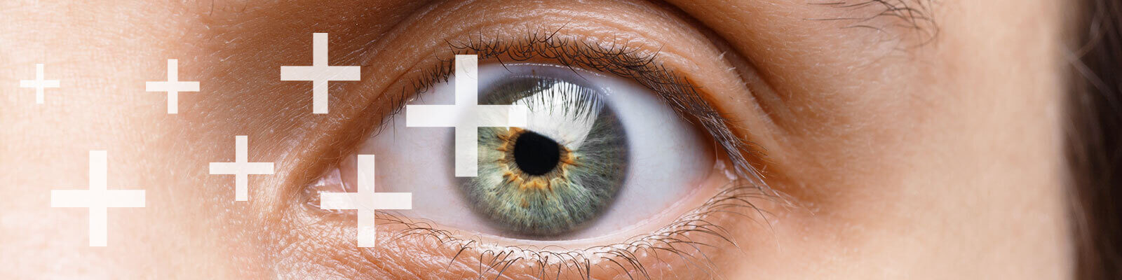
Eye and Vision Health


Article Summary
- Eye diseases can cause damage and blindness if not treated soon enough.
- The most common symptoms for eye diseases include blurriness of vision, discharge from the eyes, eye pain, excessive eye tearing, sense of dryness, light sensitivity, seeing flashes or floaters, or total vision loss.
- If you have any concerns about your eyes or vision, visit an ophthalmologist.
The eye is the window to the soul. Most people, however, do not know a lot about their eyes, rather than maintaining their health. It is incredible how such a small organ can impact our lives more than anything.
Especially with how common blinding diseases are around us. Today, let us learn more about the two most precious jewels in our faces, to see how we can care for them, and when to seek advice whenever we have an eye problem.
Eye Health
There are many possible symptoms of eye disease. If you have any concerns about your eyes or vision, visit an ophthalmologist.
A complete medical eye exam by an ophthalmologist could be the first step toward saving your sight. But first, let us learn some terminology to understand the different parts of the eye, to best describe them.
Eye Anatomy
Cornea
The cornea is in the front most part of the eye. It is the clear, dome-shaped covering in front of the iris and pupil. It’s naturally transparent and acts as a lens covering the colored part of your eyes, also known as the iris. It is precisely like the glass part on the face of your watch.
The cornea serves as a powerful refractive lens, without which your eye will be remarkably shortsighted. The cornea should always be clear and transparent; therefore, you may notice that no blood vessels are seen traveling through the cornea. There are vessels around it, though, in the white part of our eyes known as the conjunctiva.
Nevertheless, the cornea receives its nutrition directly from oxygen in the air or other nutritious fluids inside your eye. If a foreign body entered the cornea, this could easily cause a corneal abrasion, or in more severe cases, corneal ulcers. These eye problems require urgent evaluation by an ophthalmologist.
Tear Ducts
Most people think that we only produce tears when we cry. But we actually make tears continuously throughout the day. Tear production is a very important physiological process to maintain the eyes moist and sterile.
Therefore, hundreds of small tear glands around the eye help with the basal tear secretion. These tears are continuously drained from the surface of the eye through two main tear ducts.
These ducts travel through the eyelids and drain their secretions into the back of the nose. That is why when you cry; you will find yourself reaching out to a napkin to blow your nose. That is also why sometimes you can feel the taste of an eye drop in your throat after you put it in.
The tear duct is also called the nasolacrimal duct. Patients who develop allergies in their eyes suffer from excessive tear secretion throughout the day, and they need to be seen by an ophthalmologist, not just to prescribe a suitable treatment for their allergies, but also to follow up on possible complications for excessive tears and itching, which may include the development of inflammatory bumps under the eyelids, also known as papillary conjunctivitis.
Iris and Pupil
The iris is the colored part of the eye. Most people have an iris with a black, brown, blue, or green color. But the iris function is not limited to being an integral part of your eye’s beauty.
It is a mighty muscle, which, when contracts it opens up the pupil (the central dark opening in the iris that allows light to enter the eye), this pupil dilation allows more light to enter into the eye and provides a wider field of vision. This happens naturally to all of us in the dark and in situations of fear, fight, or flight. The iris also plays a significant role in patients with glaucoma.
When it relaxes, it frees up more space to the drainage system inside the eye, which allows for fluids inside the eye to get drained, thereby preventing pathologic increases in eye pressure. This is a fundamental principle in patients with glaucoma who suffer from high pressure of these fluids inside their eyes.
Lens
Inside our eyes, we have a natural lens. That is different from the cornea, which is an external lens. This “internal” lens bends (refracts) light rays that come into the eye to help us see. The lens is one of the major refractive components of the eye. It is located inside the eye right behind the iris. A clear part of the eye behind the colored iris. It helps to focus light on the retina so you can see.
The lens’s function is to primarily help with accommodation, which is the ability to change the refractive power of the eye to see near objects when increased, and far objects when decreased. This function is achieved by contraction of a particular muscle surrounding the lens.
Diseases involving the lens primarily include cataracts, which simply means clouding and opacity of the lens. Cataracts can occur due to a variety of reasons, chief among which is simply old age.
Retina
As for the retina, the best way to describe it is to think of the eye as we think of old cameras. The cornea and the lens would be together with the camera’s front lens, and the retina would be the film inside the camera where all the pictures are captured.
This means that the retina is the main neurosensory component of the eye. It is the light-sensitive tissue lining the back of the eye.
The macula is a small area of the retina responsible for your central, detailed vision. And it is considered a natural extension of the brain. It can be damaged in a variety of diseases, including diabetic retinopathy, age-related macular degeneration, retinal vessel occlusion, and hypertensive retinopathy. All these vision problems require a brief eye exam by an ophthalmologist, and regular follow up visits by retina specialists.
Optic Nerves
The optic nerve is what transmits all the signals from the eyes of the brain. It transmits all the images we see and translates them into neurological signals, which then the brain translates into shapes, colors, and faces.
In many cases, the optic nerve can get inflamed due to a variety of immune diseases, including lupus, sarcoidosis, and rheumatoid arthritis. It is also affected more commonly in patients with glaucoma, where parts of it die and become atrophic.
Common Eye Complaints
While numerous diseases can affect the eye and vision, only a handful of complaints the patient may feel. That is because lots of eye diseases can actually keep progressing in silence without the patient noticing any difference.
And by the time the patient starts complaining and noticing that their vision is not the same way it was, diseases are usually advanced, and not much can be done to help stop the progression or reverse the damage. That is why regular eye exams are crucial.
The most common symptoms for eye diseases include blurriness of vision, discharge from the eyes, eye pain, excessive eye tearing, sense of dryness, light sensitivity, seeing flashes or floaters, or total vision loss.
Refractive Errors
Refractive error is when the eye does not bend (refract) light properly. Light does not focus correctly, so images are not clear. These include nearsightedness (myopia), farsightedness (hyperopia), astigmatism, and presbyopia.
In myopia, close objects look clear but distant objects appear blurry. Astigmatism is an imperfection in the curvature of your eye’s cornea or lens. Presbyopia is when your eyes gradually lose the ability to see things clearly up close.
These errors are generally corrected using eyeglasses. They can also be corrected with contact lenses or refractive surgery such as LASEK.
Glaucoma
Glaucoma is a prevalent eye condition. It is a disease that affects the optic nerve, which connects the eye to the brain. If your eye does not circulate fluid inside the eye properly, pressure builds inside the eye and damages the optic nerve.
Glaucoma constitutes a restriction of the field of vision, mainly peripherally. It also includes atrophy to the optic nerve, and in many patients, the elevation of the pressure inside the eye.
Cataract
A cataract is the opacification of the lens inside the eye. It is the most common indication for eye surgery. It is expected for everyone to develop cataracts if they live long enough, as it is a natural aging-related change in the lens’s chemical components.
If you have a cataract, your lens has become cloudy. It is like looking through a foggy or dusty car windshield. Things look blurry, hazy, or less colorful with a cataract. It is usually treated through cataract surgery.
AMD
Age-related macular degeneration is one of the most common causes of blindness in the western world. It involves progressive atrophy of the light receptors in the retina, accumulation of lipid deposits, and fibrous materials in the retina’s deep layers. Some patients also formed new blood vessels that can bleed easily inside the eye and cause severe vision loss.
AMD causes visual loss, mainly in the central field of vision. You cannot see fine details, whether you are looking at something close or far. For instance, imagine you are looking at a clock with hands. With AMD, you might see the clock’s numbers but not the hands.
Amblyopia
Amblyopia is an irreversible loss of function of 1 of the eyes. It usually happens in children with a squint, where 1 of the eyes does not see very well due to misalignment, and thereby the child would prepare to use the other eye and learn to ignore using the squinting eye.
In these cases, the squinting eye starts losing its function, and the brain learns to disregard all the signals coming from this eye, with time, the connection between this eye and the brain is lost completely and irreversibly. Amblyopia is also known as the lazy eye.
Diabetic Retinopathy
Diabetic retinopathy is the earliest complication of diabetes. It usually occurs earlier than diabetic nephropathy and diabetic neuropathy. It involves the development of new blood vessels, small aneurysms, microscopic bleeding, swelling of the retinal layers, and in the late stages, significant bleeding inside the eye.
Diabetic retinopathy requires regular follow-up with an ophthalmologist. It is also recommended that all people with diabetes receive a dilated fundus examination by an ophthalmologist every 6 to 12 months.
Retinal Detachment
The retina is composed of 10 different layers. When these layers get separated from each other, either because of trauma, diabetes, or age-related changes, this separation is called retinal detachment. This is usually manifested by seeing floaters, seeing flashes of light, or sudden loss of vision usually described as seeing a curtain falling in front of the patient.
Dry Eye Syndrome
Dry eyes are, by far, a widespread problem in the western world. It is more common in weathers with low humidity in the air, and in families with a history of dry eye disease. It is usually a combination of deficiency and secretion of the tears, and excessive drainage of these tears causes deficient lubrication of the eye’s surface.
Our eyes need tears to stay healthy and comfortable. If your eyes do not produce enough tears, it is called dry eye. Dry eye is also when your eyes do not make the right type of tears or tear film.
Color Blindness
Color blindness occurs when you are unable to see colors in a normal way. It is also known as color deficiency. Color blindness often happens when someone cannot distinguish between specific colors. This usually happens between greens and reds, and occasionally blues. In the retina, two types of cells detect light.
They are called rods and cones. Rods detect only light and dark and are very sensitive to low light levels. Cone cells detect color and are concentrated near the center of your vision. Three types of cones see color: red, green, and blue. The brain uses input from these cone cells to determine our color perception.
Diabetic Macular Edema
Macular edema happens when fluid builds up on the retina and causes swelling and blurry vision. Diabetes can cause macular edema.
Too much blood sugar can damage the tiny blood vessels in the retina or blocked them completely. This damage or blockage causes leaking or oozing of fluid and blood into the retina. This fluid can cause swelling or edema, which can cause vision problems or blindness.
Ocular Hypertension
Ocular hypertension is when the pressure inside the eye (intraocular pressure or IOP) is higher than normal. With ocular hypertension, the front of the eye does not drain fluid properly. This causes eye pressure to build up. Higher than normal eye pressure can cause glaucoma. Glaucoma is a disease where eye pressure damages the optic nerve, causing vision loss.
Ocular hypertension is not the same as glaucoma. With ocular hypertension, the optic nerve looks normal, and there are no signs of vision loss. However, people with ocular hypertension are at increased risk for glaucoma and are considered “glaucoma suspects.”
Tips for Eye Health
- Try to get a comprehensive dilated eye exam. This is a simple, affordable, and painless physical examination that you can get at your optometrist or ophthalmologist office. It is recommended for everyone above the age of 40 to get a regular eye exam every year. This can early detect lots of eye diseases that take years to show symptoms.
- Protect your eyes from the damaging effect of the sun. The use of sunglasses of good quality that blocks ultraviolet rays has been proven to help against multiple eye diseases, including macular degeneration.
- Take regular rests from reading and using computer screens by applying the 20, 20, 20 rule. Take a break every 20 minutes, look at something 20 feet away, for 20 seconds before you get back to work.
- If you use contact lenses, wash your hands with soap and water before you take them out or put them in. Be sure to never sleep with them. And always replace them and never leave them and beyond their intended duration of use set by the manufacturer. Any form of carelessness when it comes to contact lenses can have severe and unforgiving consequences. Sleeping in contacts or using saliva or water as a wetting solution, or using expired solutions can all result in deep and infected corneal ulcers that can cause irreversible visual loss.
- Following healthy habits, such as a nutritious diet, simple exercises, and maintaining a healthy lifestyle, can extend to benefit your eyes and ocular health.
- Smoking is one of the most famous risk factors for multiple eye diseases, including cataracts and macular degeneration. It can also cause significant damage to the optic nerves.
- Try to include dark leafy green vegetables in your diet. Produce like spinach, kale, collard greens, and carrots are excellent sources of nutrition to the eyes. Meat products that are high in omega-3 fatty acids, such as salmon, tuna, and halibut are also of great importance.
- Find out if you have a family history of eye diseases. Many eye problems tend to be genetic and run in families. These include conditions such as cataracts, diabetic retinopathy, age-related macular degeneration, and glaucoma. If you have these diseases in the family, you may want to get a comprehensive eye exam earlier than most people because you may be at increased risk.
This article is for informational purposes only and does not serve as medical advice. The details provided here are not a replacement for, and should never be depended upon as, professional medical advice. Always consult your physician regarding the potential risks and benefits of any treatment.
Our Medical Review Process
At Ben’s Natural Health, we are committed to maintaining the highest standards of accuracy, transparency, and scientific integrity. Every piece of content is carefully developed by medical professionals and undergoes a thorough review every 12 to 24 months. This ensures that our information remains current, reliable, and rooted in credible, evidence-based research. We reference only peer-reviewed studies from reputable medical journals, providing full citations and direct links to enhance trust and confidence. Learn more about our medical review process and research standards.
Our Editorial Guidelines
For over 25 years, Ben’s Natural Health has been a trusted source of scientifically backed, reliable health information. Our editorial guidelines uphold the highest quality and integrity for every article we publish. Each piece is written by qualified experts and undergoes independent quality checks. We prioritize transparency by clearly displaying contributor credentials and biographies at the beginning of every article. Read more about our editorial standards.
Medical Disclaimer
The content on this blog is for informational purposes only and should not be considered a substitute for professional medical advice, diagnosis, or treatment. While our articles are authored and reviewed by licensed medical professionals, they may not address your specific health concerns. Always consult a qualified healthcare provider before making any medical decisions.
Article Update History
Updated on 27 May, 2026 (Current Version)
Created on 14 August, 2020
Explore More
How To Grow Your Beard Faster and Thicker?
The process of growing hair, whether on your head or face, can be long and quite frustrating. A lot of different factors can affect ...